
| name | Amanita ifeifensis | ||||||||||||
| author | Tulloss, S. D. Russell & Alofe | ||||||||||||
| name status | nomen provisorum | ||||||||||||
| english name | "Ife-Ife Ringless Amanita" | ||||||||||||
| etymology | Ife-Ife + -ensis, occuring in | ||||||||||||
| GenBank nos. |
Due to delays in data processing at GenBank, some accession numbers may lead to unreleased (pending) pages.
These pages will eventually be made live, so try again later.
| ||||||||||||
| intro |
Olive text indicates a specimen that has not been
thoroughly examined (for example, for microscopic details) and marks other places in the text
where data is missing or uncertain. The following material is based on original research of R. E. Tulloss. Molecular data are courtesy Stephen D. Russell, Purdue University. | ||||||||||||
| stipe | universal veil as saccate volva, membranous, tubular, apparently attach to lower sides of stipe (not just at base). | ||||||||||||
| odor/taste | record not available. | ||||||||||||
| macrochemical tests |
record not available. | ||||||||||||
| hymenial trama | bilateral, divergent. | ||||||||||||
| subhymenium | probably immature in this material, shallow, perhaps only one layer of inflated cells below longest basidia, nearly subparenchymtous in some regions of section, with clear branching structure and less inflation of cells in other regions. | ||||||||||||
| basidia | 41 - 56 × 9.5 - 10.5 μm, 4-sterigmate, arising from branching structure of inflated cells and short uninflated cells [note: material seems not completely mature.—ed.]; clamps very rare if present. | ||||||||||||
| universal veil | On pileus: with plentiful gelatinized elements still clearly intimately attached to pileipellis. | ||||||||||||
| stipe context | longitudinally acrophysalidic; dominantly hyphal at surface, dominantly acrophysalidic in context; filamentous undifferentiated hyphae 1.9 - 7.6 μm wide, partially gelatinized near stipe surface, branching; acrophysalides 152 - 292 × 20 - 36 μm; vascular hyphae 2.6 - 13.7 μm with insoluble content, relatively common, especially near surface, scattered, rarely branched; clamps rare if present (not observed). | ||||||||||||
| lamella edge tissue | sterile; subparenchymatous in side view; filamentous undifferentiated hyphae infrequent on surfaces; inflated cells in ca. 7 layers, thin-walled or with wall up to 0.5 μm thick, 13.5 - 28 × 10.0 - 27 μm, globose to pyriform to subglobose to ellipsoid, in chains of up to six or more. | ||||||||||||
| basidiospores | RET: [40/1/1] (10.4) 11.3 - 12.6 (-15.0) × (5.2-) 5.3 - 6.0 (-7.5) μm, (L = 12.0 μm; W = 5.8; Q = (1.39-) 1.91 - 2.26 (-2.40); Q' = 2.09), hyaline, colorless, thin-walled, smooth, inamyloid, elongate to cylindric (rarely ellipsoid), adaxially flattened; apiculus sublateral, elongate to cylindric, occasionally constricted, occasionally expanded at one end; contents monoguttulate, with or without additional small granules; color in deposit not recorded. | ||||||||||||
| ecology | Associated with Brachystegia eurycoma (Fabaceae subgenus Caesalpinioideae), a mycorrhizal leguminous tree. | ||||||||||||
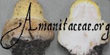
| material examined | NIGERIA: OSUN STATE—Ife-Ife, Obafemi Owolowo State Univ., Parks and Gardens Unit, 27.x.2003 F. V. Alofe #2 (RET 329-7, nrITS & nrLSU seq'd.) | ||||||||||||
| discussion |
In evaluating the microscopic data, it should be noted
that only one exsiccatum was available and that it
was not fully mature. Hence, for example, the
spores and the lamella trama were viewed in sections
taken near the stipe-end of a lamella—the
most mature area. Whether these obsevations
would be identical to those from more mature material
is not clear. The nrLSU 5' motif (TCTGACCTCAAATCA...) places this species in the provisional "subsection Penetratrices". Among the nearly sixty species with this motif, the present one is the only one known to bear spores with Q' > 2.0. The only other species in the Penetratrices with a techtab (Amanita sp-N58) has a maximum Q value of 1.81. Unfortunately because of difficulties with original documentation, it is not possible to connect this collection with more macroscopic information than can be deduced from the exsiccatum. To this we can add both anatomical data from my 2003 examination of the specimen and rDNA sequence data. | ||||||||||||
| citations | —R. E. Tulloss | ||||||||||||
| editors | RET | ||||||||||||
Information to support the viewer in reading the content of "technical" tabs can be found here.

Text and User-Generated Sporographs are published under the Creative Commons License.

In the case of a taxon page, image credits are on the 'image' tab.
