
| name | Amanita virella |
| name status | nomen acceptum |
| author | E-J. Gilbert ex Singer |
| english name | "Beeli's Green-Staining Amanita" |
| intro |
The following is based on the original description by Beeli (1935) as well as the description by Gilbert (1941). |
| cap |
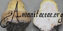
The cap of A. virella is 50 - 60 mm wide, convex at first, then plano-convex, at first dark umbre, then becoming greenish umbrinous, fleshy, with a slightly striate margin. The volva is present as friable thick pyramidal warts or as small narrow warts mainly over the center. The flesh is 3 - 5 mm thick above the stem, firm, whitish cream, becoming intense green. |
| gills |
The gills are free, yellowish white to pale sulfur yellow, then becoming pale green, 4 - 5 mm broad, pointed at both ends. |
| stem |
The stem is 90 - 120 × 5 - 12 mm, cylindric, pallid olivaceous to grayish, solid, smooth, with a bulbous base that is 15 - 20 mm wide and subglobose to ellipsoid. The ring is placed high on the stem, membranous, persistent, skirt-like, off-white, and dark umber on the upper surface at the edge. The volva is friable and leaves only a pale brown circlet at the base of the stem above the bulb. The flesh is white, becoming green near the top of the stem. |
| odor/taste |
The odor is penetrating. The taste is bitter. |
| spores |
Spores from the original description by Beeli (1935) measure 10 - 11 × 6 - 7 µm and are amyloid. Spores measured from Gilbert's spore drawings (1940) measure 9.1 - 11.4 × 6 - 8 µm and are broadly ellipsoid to ellipsoid. |
| discussion |
This species was originally described from the former Belgian Congo where it occurred singly in dry forest. Gilbert emphasized that the green staining reaction was notable for its intensity. Gilbert gives additional information about this species but provides no source. His observations are not justified by the original description or the watercolor provided to Beeli by Madame Goossens. In this additional material, he says that the flesh in the stem can have a yellowish tint at first and may become pinkish at the top of the stem before becoming green. Anyone able to provide us information about the source of Gilbert's observations will receive our sincere gratitude.—R. E. Tulloss and L. Possiel |
| brief editors | RET |
| name | Amanita virella | ||||||||
| author | E.-J. Gilbert ex Singer. 1951 ["1949"]. Lilloa 22: 38. | ||||||||
| name status | nomen acceptum | ||||||||
| english name | "Beeli's Green-Staining Amanita" | ||||||||
| synonyms |
≡Amanita virella E.-J. Gilbert nom. inval. 1941. Iconogr. Mycol. (Milan) 27, suppl.: 384, tab. 62. [New name not definitely accepted by author. ICBN §34.1(a)]
≡Amanita virescens Beeli nom. illeg. 1931. Bull. Soc. Roy. Bot. Belgique 63: 106, pl. 9 (fig. 11). [Posterior homonym. ICBN §53.1]
≡Aspidella virescens (Beeli) E.-J. Gilbert. 1940. Iconogr. Mycol. (Milan) 27, suppl. (1): 79, tab. 51 (fig. 5). non Amanita virescens Pers. nom. dub. 1801. Syn. Meth. Fung. 2: 255. The editors of this site owe a great debt to Dr. Cornelis Bas whose famous cigar box files of Amanita nomenclatural information gathered over three or more decades were made available to RET for computerization and make up the lion's share of the nomenclatural information presented on this site. | ||||||||
| MycoBank nos. | 560196, 163586, 284335 | ||||||||
| GenBank nos. |
Due to delays in data processing at GenBank, some accession numbers may lead to unreleased (pending) pages.
These pages will eventually be made live, so try again later.
| ||||||||
| holotypes | BR (implicit) | ||||||||
| revisions | Bas. 1969. Persoonia 5: 568. [discussion of status] | ||||||||
| selected illustrations | Beeli. 1935. Fl. Champ. Congo 1: pl. 3 (fig. 5). | ||||||||
| intro |
The following text may make multiple use of each data field. The field may contain magenta text presenting data from a type study and/or revision of other original material cited in the protolog of the present taxon. Macroscopic descriptions in magenta are a combination of data from the protolog and additional observations made on the exiccata during revision of the cited original material. The same field may also contain black text, which is data from a revision of the present taxon (including non-type material and/or material not cited in the protolog). Paragraphs of black text will be labeled if further subdivision of this text is appropriate. Olive text indicates a specimen that has not been thoroughly examined (for example, for microscopic details) and marks other places in the text where data is missing or uncertain. The following material is derived from the protolog of the present taxon, (Beeli 1935), (Gilbert 1940 & 1941), and (Bas 1969). | ||||||||
| pileus |
from protolog: 50 - 60 mm wide, brown to fuligineous, plano-convex; context fleshy, firm, white, becoming green in upper part of disc; margin substriate; universal veil as covering of warts and squamules, dark. Bas (1969): margin subsulcate, nonappendiculate. | ||||||||
| lamellae | from protolog: free, density not described, yellowish white, then becoming green, 4 - 5 mm broad. | ||||||||
| stipe | from protolog: 90 - 120 × 5 - 12 mm, grayish, cylindric, smooth; bulb subabrupt and submarginate (per figure); context solid, firm, white; partial veil superior, membranous, pendent; universal veil as rings on and just below bulb margin (per figure), otherwise ephemeral. | ||||||||
| odor/taste | from protolog: Odor acrid. Taste bitter. | ||||||||
| macrochemical tests |
none recorded. | ||||||||
| pileipellis | Beeli (1935): filamentous hyphae broad; "excretory" (vascular?) hyphae present. | ||||||||
| pileus context | not described. | ||||||||
| lamella trama | not described. | ||||||||
| subhymenium | not described. | ||||||||
| basidia | Bas (1969): 2-sterigmate (in type); clamps lacking. | ||||||||
| universal veil | not described. | ||||||||
| stipe context | not described. | ||||||||
| partial veil | not described. | ||||||||
| lamella edge tissue | not described. | ||||||||
| basidiospores |
from protolog: 10 - 11 × 6 - 7 μm, hyaline, smooth, ellipsoid. [Note: No sporograph generated.—ed.] Beeli (1935): white in deposit. Gilbert (1940 & 1941): [4/1/1] 9.1 - 11.4 × 6.0 - 8.0 μm, (L = 10.6 μm; W = 7.4 μm; Q = 1.36 - 1.52; Q = 1.43), hyaline, smooth, amyloid, ellipsoid; apiculus sublateral and subcylindric to truncate conic (all per figure); contents not described; white in deposit. [Note: Spore measurements are taken from the four drawings of (Gilbert 1940: tab. LI (fig. 5)) that are in apparent lateral view.—ed.] Bas (1969): [-/-/-] 9 - 11 × 6.5 - 8 μm, (est. Q = 1.35 - 1.45), amyloid, ellipsoid. | ||||||||
| ecology | from protolog: Scattered. On soil in dry forest. | ||||||||
| material examined |
from protolog: CONGO, DEMOCRATIC REPUBLIC OF: PROV. EQUATEUR—Territoire Lisala - Binga [2°23'41" N/ 20°25'25" E, 361 m], Beeli (1935): CONGO, DEMOCRATIC REPUBLIC OF: PROV. EQUATEUR—Territoire Lisala - Binga [2°23'41" N/ 20°25'25" E, 361 m], Gilbert (1940 & 1941): CONGO, DEMOCRATIC REPUBLIC OF: PROV. EQUATEUR—Territoire Lisala - Binga [2°23'41" N/ 20°25'25" E, 361 m], | ||||||||
| discussion |
Bas (1969: 568-569) briefly revised the type of the present species because Gilbert had placed with other taxa that are assignable to sect. Lepidella. He commented on proper placement as follows:
"Gilbert (1940: 70) place this species in his genus Aspidella, which is identical with Amanita section Lepidella in the present work, but the type material revealed a distinctly coloured pileipellis combined with a non-appendiculate, subsulcate margin of the cap, a membranous ring, a volva that leaves only fugacious remnants on the small to medium-sized bulb of the stem, and ellipsoid, amyloid spores 9 - 11 × 6.5 - 8 μ on 2-spored, clampless basidia. It is clear that A. virella belongs to Amanita section Validae." | ||||||||
| citations | —R. E. Tulloss | ||||||||
| editors | RET | ||||||||
Information to support the viewer in reading the content of "technical" tabs can be found here.
| name | Amanita virella |
| bottom links |
[ Keys & Checklists ] |
| name | Amanita virella |
| bottom links |
[ Keys & Checklists ] |
Each spore data set is intended to comprise a set of measurements from a single specimen made by a single observer; and explanations prepared for this site talk about specimen-observer pairs associated with each data set. Combining more data into a single data set is non-optimal because it obscures observer differences (which may be valuable for instructional purposes, for example) and may obscure instances in which a single collection inadvertently contains a mixture of taxa.


Text and User-Generated Sporographs are published under the Creative Commons License.

In the case of a taxon page, image credits are on the 'image' tab.