
| name | Limacellopsis sp-Burnett-5-xi-1994 |
| name status | cryptonomen temporarium |
| author | Tulloss |
| images |
, U.S.A.") 1. Limacella cf. roseicremea, Lummi Island, Whatcom County, Washington (state), U.S.A. |
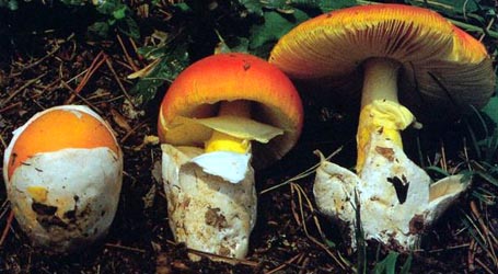
| cap | The cap is white to pale cream with a pink tint and, eventually, is irregularly flattened. The cap margin is incurved at first and not striate. A gluten layer is present. |
| gills | The gills are probably free, close, and whitish. |
| stem | The stem is pallid and dry. In dried material, the stipe has a distinct, subglobose bulb. The stem's flesh is solid, with a distinct, sordid central cylinder in dried material. The stem has a superior, white, skirt-like annulus that points upward at first like a funnel; after drooping, the ring preserved a groove around the stem when viewed from above. |
| odor/taste | The odor and taste were not reported by the collector. |
| spores | The spores measure (4.1–) 4.3–6.0 (–7.2) × (3.2–) 3.4–4.6 (–5.5) µm and are broadly ellipsoid to ellipsoid and mostly inamyloid, with a few dextrinoid. Clamps are plentiful at bases of basidia. |
| discussion | This entity clearly belongs in Limacellopsis . The only North American candidate in that section that is similar to Ms. Burnett's material is Limacella roseicremea. The only reason that there is any hesitation about identifying the present collection as L. roseicremea is the very minimal available information about the latter.—R. E. Tulloss |
| brief editors | RET |
| name | Limacellopsis sp-Burnett-5-xi-1994 | ||||||||
| author | Tulloss | ||||||||
| name status | cryptonomen temporarium | ||||||||
| GenBank nos. |
Due to delays in data processing at GenBank, some accession numbers may lead to unreleased (pending) pages.
These pages will eventually be made live, so try again later.
| ||||||||
| intro |
Olive text indicates a specimen that has not been
thoroughly examined (for example, for microscopic details) and marks other places in the text
where data is missing or uncertain. The following material is based upon original research by R. E. Tulloss. | ||||||||
| pileus | ??, white to pale cream with pink tint, ??, eventually irregularly planar; context ??; margin nonstriate, incurved at first, ??; gluten layer present. | ||||||||
| lamellae | ??, close, white or whitish; lamellulae ??. | ||||||||
| stipe | ??, dry; bulb subglobose and distinct in exsiccata; context solid (distinct central cylinder filled with sordid tissue in exsiccata); partial veil superior, white, skirt-like, membranous, at first infundibuliform, retaining sinus at stipe after drooping (as, e.g., in L. guttata), ??; gluten layer ??. | ||||||||
| odor/taste | not recorded. | ||||||||
| macrochemical tests |
none recorded | ||||||||
| pileipellis | absent | ||||||||
| lamella trama | bilateral; central stratum distinct, comprising interwoven filamentous undifferentiated hyphae (?? µm wide) with frequent fusiform intercalary segments (?? × ?? µm ); ??. | ||||||||
| basidia | 26–30 × 6.3–7.8 µm, 4-sterigmate, with sterigmata up to 3.8 × 1.5 µm; clamps prominent, plentiful. | ||||||||
| gluten layer | On pileus: filamentous hyphae supporting gluten pile 1.7–6.0 µm wide, branching, gelatinizing extensively in older material; terminal cells approximately conic to pyriform to fusiform to constricted-subfusiform to "sausage-shaped," occasionally capitulate, frequently rostrate, 12.5–49 × 4.0–13.5 µm, 2.0–5.5 µm wide at base, with length/max.-width ratio = 2.1–6.0, occasionally with pale yellow vacuolar pigment, erect at first, then collapsing without dominant orientation and gelatinizing, arising either from uninflated or slightly inflated hyphal segments or from subglobose to broadly ellipsoid inflated cells (10.8–42 × 9.5–33 µm, apparently sometimes in short chains); clamps common. On stipe: ??. | ||||||||
| stipe context | longitudinally acrophysalidic; filamentous undifferentiated hyphae 1.9–8.5 µm, dominating, branching; acrophysalides 40–92± × 7.8–20 µm; vascular hyphae not observed??; clamps common. | ||||||||
| lamella edge tissue | fertile | ||||||||
| basidiospores | RET from proposed epitype: [60/3/1] (4.1–) 4.3–6.0 (–7.2) × (3.2–) 3.4–4.6 (–5.5) µm, (L = 5.0–5.2 µm; L’ = 5.2 µm; W = 3.9–4.2 µm; W’ = 4.0 µm; Q = (1.06–) 1.09–1.53 (–1.94); Q = 1.26–132; Q’ = 1.30), hyaline, colorless, smooth, dominantly inamyloid, occasionally dextrinoid, broadly ellipsoid to ellipsoid, infrequently subglobose, rarely elongate, adaxially flattened; apiculus sublateral, cylindric; content granular to mono- to multiguttulate; color in deposit not recorded. | ||||||||
| ecology | In small groups. | ||||||||
| material examined | U.S.A.: WASHINGTON—Whatcom Co. - Lummi Isl., 5.xi.1994 Nancy Burnett s.n. (RET 136-9, nrITS & nrLSU seq'd.). | ||||||||
| discussion |
This collection matches the original description of L. roseicremea except for the size and shape of the spores. However, the few spores that I was able to find on a surviving paratype of L. roseicremea are a good match to the spores of the present fungus. For purposes of comparison, the available spore data (red figure) from the type of L. roseicremea is given here: [8/1/1] 4.5 – 6.2 × 3.5 – 4.5 µm, (L = 4.8 µm; L’ = 4.8 µm; W = 3.7 µm; W’ = 3.7 µm; Q = (1.21–) 1.22 – 1.43; Q = 1.30; Q’ = 1.30). For purposes of comparison, RET's spore data (green figure) from European material of L. guttata is given here: [12/1/1] 4.8 - 5.8 (-6.1) × (3.9-) 4.0 - 4.3 (-5.0) µm, (L = 5.3 µm; W = 4.1 µm; Q = 1.20 - 1.42; Q' = 1.30). For purposes of comparison, the very limited available spore data (orange figure) of L. solidipes is given here: [-/-/-] 4 – 5 × 4 – 5 µm, (est. Q = 1.0 – 1.25). [Nota: This data should not be presumed to be reliable.—ed.] The comparison of sporographs (above) indicates that, in the present state of knowledge, spore size and shape do not provide a sufficient data set for segregation the taxa reported from North America that are similar to L. roseicremea. Hence, more detailed microscopic studies, including revision of all relevant types will be necessary to develop clear species concepts in this group of taxa within Limacella sect. Amanitellae. It is possible that some of the names will prove to be taxonomic synonyms. The terminal cells of the gluten supporting hyphae in the pileal gluten layer conform very closely to the terminal cells in the corresponding tissue of L. guttata; as described by Neville and Poumarat (2004). Moreover, a persistent, membranous partial veil is clearly present. Therefore, it is proposed that the present species should be considered to belong in Limacella subsect. Amanitellae. | ||||||||
| citations | —R. E. Tulloss | ||||||||
| editors | RET | ||||||||
Information to support the viewer in reading the content of "technical" tabs can be found here.
| name | Limacellopsis sp-Burnett-5-xi-1994 |
| bottom links | [ Keys & Checklists ] |
| name | Limacellopsis sp-Burnett-5-xi-1994 |
| bottom links | [ Keys & Checklists ] |
Each spore data set is intended to comprise a set of measurements from a single specimen made by a single observer; and explanations prepared for this site talk about specimen-observer pairs associated with each data set. Combining more data into a single data set is non-optimal because it obscures observer differences (which may be valuable for instructional purposes, for example) and may obscure instances in which a single collection inadvertently contains a mixture of taxa.


Text and User-Generated Sporographs are published under the Creative Commons License.

In the case of a taxon page, image credits are on the 'image' tab.